Neeraj Awasthy, Premila Pul.
Department of Pediatrics, Safdarjung Hospital, Delhi, India.
ADDRESS FOR CORRESPONDENCE
Dr Neeraj Awasthy, 123, Nandkunj, Vikaspuri, New Delhi- 10018, India.
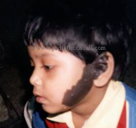
Email: n_awasthy@yahoo.com | | Abstract | | Epidermal nevi syndrome (ENS) is a multisystem neurodermatological disorder in which epidermal nevi may be associated with neurological, skeletal, ocular and other cutaneous anomalies as well as malignancies with potentially devastating consequences. Most cases are sporadic though occasionally autosomal dominant transmission has been documented. The objective of the present communication is to sensitize regarding the presence of the entity. | | | | Case Report | A 4 year old girl, resident of Delhi presented with unilateral non-progressive black discoloration of left cheek since birth and history of recurrent generalized seizures since 3 months of age. There was no history of drug intake, trauma or neurological insult. There was no family history of seizure disorder or similar lesions. Examination of the child revealed a large papular area dark blue in color involving the left cheek and pinna, with well defined margins (Fig.1). Neurological system examination was within normal limits. All other systemic examination was within normal limits. Investigations showed a normal hematological profile, with normal liver and renal function tests. EEG was suggestive of diffuse cortical seizure activity; CECT was within normal limits. On biopsy the lesion was described as florid epidermal hyperplasia with acanthosis and hyperkeratosis. Patient was managed conservatively with antiepileptic drugs in appropriate dosage with regular follow up.
Figure 1:
 | | | | Discussion | Epidermal nevi are hamartomas that are characterized by hyperplasia of the epidermis and adenexal structures. These have been estimated to occur in 1:1000 live births, affecting the sexes equally (1). They occur sporadically, however familial cases have been reported. These have classified into a number of distinct variants based on the extent of involvement, clinical morphology and the predominant epidermal structure of the lesion. Typically, epidermal nevi are present at birth or early infancy but have been described to appear in puberty. In one of the series of cases, 60% of lesions were present at birth, 80% were found by the end of the first year of life, and most of the remainder developed between the ages of 1 and 7 years (1).
Most common site reported for these lesions is on the head and neck region (2). Lesions may enlarge slowly in childhood, become darker and thicker, and by adolescence reach a stable size after which further growth is unlikely.
Epidermal nevi are organoid nevi arising from the pluripotential germinative cells in the basal layer of the embryonic epidermis. These cells give rise to not only keratinocytes but also to skin appendages. These nevi have often been classified according to the predominant component, resulting in the term nevus verrucosus (keratinocytes), nevus sebaceous (sebaceous glands), naevus comedonicus (hair follicles), and nevus syringocystadenoma papilliferus (apocrine glands). However very few lesions are exclusively of one type (3). The predominant tissue may vary with the evolution of the lesion and different areas of the same lesion may show a variety of components at the same time.
ENS, also reported as Schimmelpenning syndrome or Feuerstein and Mims syndrome, refers to a disease complex consisting of epidermal nevi with developmental abnormalities of the integumentary, ophthalmologic, nervous, skeletal, cardiovascular, urogenital systems and associated malignancies. Feuerstein and Mims first described the syndrome in 1962 and the complex was entitled epidermal nevus syndrome by Solomon in 1968 (4).
Cutaneous changes, besides the epidermal nevi were found in one-third of the cases of ENS. Cafe-au-lait spots were the most common in one of the series. Hemangiomas and pigmentary changes are reported in 10-20% of the case. Skeletal changes have ranged from 15 to 70%, kyphoscoliosis being the most common. Neurological abnormalities occur in 15% to 50% of reported cases. Mental retardation, developmental delay and the seizures are the most common neurologic findings. A major concern in these cases is the neoplastic potential not only of the skin but also of the other organ systems. Basal cell carcinoma being the commonest, systemic malignancies such as Wilm's tumor, nephroblastomas, salivary gland adenocarcinoma, oesophageal and stomach carcinomas, breast carcinomas, astrocytomas and mandibular ameloblastomas have been reported to occur with higher frequency an earlier age in patients with ENS (5).
The treatment of verrucous epidermal nevi is excision, however it may not be a practical advice in extensive lesions. Alternative therapies include electrofulguration, liquid nitrogen cryotherapy, dermabrasion, intra-lesional corticosteroids and chemical peels. These modalities remove only the superficial portions of the nevus and typically give partial response. Topical therapy with podophyllin, retinoic acid, anthralin, alpha hydroxy acids are usually ineffective or provide partial response. Carbon dioxide laser treatment has been tried with efficacy in such lesion. | | | | Compliance with Ethical Standards | | Funding None | | | | Conflict of Interest None | | |
- Losee EJ, Sereletti MJ, Penniro RP Epidermal nevi syndrome: A review and case report. Ann. Plastic Surg. 1999,44(2);211-214. [CrossRef]
- Rogers M. Mc Cossin I , Commens C. Epidermal nevi and the epidermal nevus syndrome. A review of 131 cases. J Am Acad. Dermatol. 1989;20 (3):476-488. [CrossRef]
- Solmon LM, Esterly NB Epidermal and other organoid nevi. Cur Probl Pediatr 1975;6:1-56. [CrossRef]
- Paller AS. Epidermal Nevus Syndrome. Neurol Clin. 1987;5(3);451-457. [CrossRef]
- Fitzpatrick TB. In Dermatology in general medicine. 4th edition vol1. New York. Mc Graw Hill, 1993:858-864.
|
| Cite this article as: | | Awasthy N, Pul P. EPIDERMAL NEVI SYNDROME. Pediatr Oncall J. 2005;3: 49. |
|