Leonor Aires Figueiredo1, Ricardo Domingos Grilo2, Patrícia Romão2, Sónia Antunes3
1Pediatric Department, Hospital do Espírito Santo de Évora, Évora, Portugal, 2Pediatric Department, Departamento da Saúde da Mulher e da Criança, Hospital do Espírito Santo de Évora, Évora, Portugal, 3Neonatology Unit, Pediatric Department, Hospital do Espírito Santo de Évora, Évora, Portugal
Address for Correspondence: Leonor Aires Figueiredo, Largo do Sr. da Pobreza, 7000-811 Évora.
Email: leonor.figueiredo12@gmail.com
|
Question :Term newborn, male, with no relevant family history. Monitored pregnancy, prenatal ultrasounds without described alterations, early neonatal period without intercurrences. Fall of the umbilical stump occurred on the 10 th day of life.
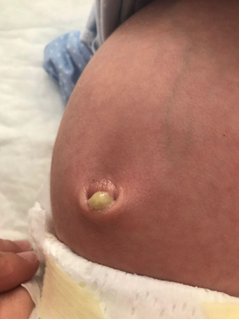
Observed on the 15 th day of life for purulent umbilical drainage, abdominal distention and maternal awareness of irritability with two days of evolution, without fever, change in intestinal transit or food refusal. Physical examination showed good vitality, a distended and tympanized abdomen with discomfort on palpation, which improved after defecation and the presence of purulent umbilical exudate, with no fetid odor and no other local inflammatory signs present. The analytical evaluation did not reveal elevation of inflammatory parameters or other alterations. A cultural examination of the exudate was performed and the patient was discharged with indication for hygiene care and surveillance. Two days later, the infant was reassessed for the reappearance of umbilical drainage with the same characteristics, with the objective examination and analytical evaluation overlapping. On suspicion of omphalitis, intravenous antibiotic therapy was started during hospitalization, with flucloxacillin 100 mg/kg/day and gentamicin 4 mg/kg/day. In the ultrasound a tubular structure was visualized between the bladder and the umbilicus, with hypoechogenicity in the umbilical region, compatible with urachal sinus. The cultural examination of the umbilical exudate revealed the presence of methicillin-sensitive Staphylococcus aureus, so gentamicin was suspended on the 3 rd day of hospitalization, having completed 10 days of intravenous flucloxacillin. There was a favorable clinical evolution, without the presence of purulent exudate or other inflammatory signs since the 3rd day of hospitalization.
The infant was discharged for a Pediatric Surgery consultation. Since then, he has remained asymptomatic under regular imaging control. The last imaging evaluation, at 21 months of age, revealed a slight prominence in the vesical aspect of the urachus with no visible communication with the bladder or umbilical region, nor cystic translation.
Figure 1. Umbilical discharge.  What is a urachal sinus? Are there other urachal anomalies?
|
Discussion :
The urachus or median umbilical ligament, is a fibrous tubular structure that connects the upper portion of the bladder to the umbilicus. It corresponds to a vestigial remnant of two embryonic structures: the cloaca, a cephalic extension of the urogenital sinus and the allantois, a derivative of the yolk sac. Its involution and obliteration usually occur before birth. 1,2,3
Pathologies resulting from its abnormal involution are rare and are divided into four types, according to the location of the patent portion: patent urachus (50%), urachal cyst (30%), urachal sinus (15%) and urachal diverticulum (5%). 4,5,6,7
Urachal anomalies are mostly asymptomatic, with clinical manifestations being related to the underlying anomaly and resulting from the presence of local complications, such as inflammation, infection, umbilical exudate, abdominal pain or malignancy. 8,9
A patent urachus occurs when there is a connection between the bladder and the umbilicus and can cause urine leakage at the umbilicus. A urachal cyst represents a section of the urachus that did not close, but there is not a connection between the bladder and umbilicus. It is often asymptomatic and can be only detected when an ultrasound is performed for other reasons or, in rare occasions, when the cyst becomes infected, leading to abdominal pain and distension. Urachal sinus occur when the urachus did not seal close to the umbilicus and leads to a blind ending tract from the umbilicus into the urachus. Urachal sinus may infect and present with abdominal pain and drainage of fluid. A urachal diverticulum occurs when the urachus did not seal close to the bladder and leads to a blind ending tract from the bladder into the urachus and can present with a urinary tract infection. 3,10
Clinical signs and symptoms can be confused with other abdominal and pelvic pathologies, so the diagnosis is challenging. When suspected, imaging techniques are useful in the diagnosis and a wide range is available, including ultrasonography, computed tomography, magnetic resonance and fluoroscopy studies. 3 Treatment is controversial, with most authors advocating a conservative approach in asymptomatic patients, as spontaneous closure can occur. 8,10,12
This case stands out due to the rarity of the underlying pathology and early presentation, with a high level of clinical suspicion being important for its diagnosis. The presence of voluminous purulent exudate in the absence of other local inflammatory signs and systemic or laboratory repercussions led to the consideration of this diagnostic hypothesis, allowing a positive outcome. | References : | - Fahmy M. Urachal Anomalies. In: Umbilicus and Umbilical Cord. Springer International Publishing; 2018:229-252. doi:10.1007/978-3-319-62383-2_35.
- Vescovi J, Doria C, Malfussi H. Urachal sinus in an infant: a case report. Residência Pediátrica. 2022;12(3). doi:10.25060/residpediatr-2022.v12n3-365.
- Buddha S, Menias CO, Katabathina VS. Imaging of urachal anomalies. Abdominal Radiology. 2019;44(12):3978-3989. doi:10.1007/s00261-019-02205-x.
- Riaza Montes M, Antón Eguia BT, Gallego Sánchez JA. Urachal sinus: An atypical case and review of the literature. Urol Case Rep. 2023;47:102359. doi:10.1016/j.eucr.2023.102359.
- Luo X, Lin J, Du L, Wu R, Li Z. Ultrasound findings of urachal anomalies. A series of interesting cases. Med Ultrason. 2019;21(3):294. doi:10.11152/mu-1878.
- Yu JS, Kim KW, Lee HJ, Lee YJ, Yoon CS, Kim MJ. Urachal Remnant Diseases: Spectrum of CT and US Findings. RadioGraphics. 2001;21(2):451-461. doi:10.1148/radiographics.21.2.g01mr02451.
- Sreepadma S, Rao BRC, Ratkal J, Kulkarni V, Joshi R. A Rare Case of Urachal Sinus. JOURNAL OF CLINICAL AND DIAGNOSTIC RESEARCH. 2015;9(7):PD01-PD02. doi:10.7860/JCDR/2015/13243.6185.
- Sato H, Furuta S, Tsuji S, Kawase H, Kitagawa H. The current strategy for urachal remnants. Pediatr Surg Int. 2015;31(6):581-587. doi:10.1007/s00383-015-3712-1.
- Parada Villavicencio C, Adam SZ, Nikolaidis P, Yaghmai V, Miller FH. Imaging of the Urachus: Anomalies, Complications, and Mimics. RadioGraphics. 2016;36(7):2049-2063. doi:10.1148/rg.2016160062.
- Nogueras-Ocaña M, Rodríguez-Belmonte R, Uberos-Fernández J, Jiménez-Pacheco A, Merino-Salas S, Zuluaga-Gómez A. Urachal anomalies in children: Surgical or conservative treatment? J Pediatr Urol. 2014;10(3):522-526. doi:10.1016/j.jpurol.2013.11.010.
- Naiditch JA, Radhakrishnan J, Chin AC. Current diagnosis and management of urachal remnants. J Pediatr Surg. 2013;48(10):2148-2152. doi:10.1016/j.jpedsurg.2013.02.069.
- Gleason JM, Bowlin PR, Bagli DJ, et al. A Comprehensive Review of Pediatric Urachal Anomalies and Predictive Analysis for Adult Urachal Adenocarcinoma. Journal of Urology. 2015;193(2):632-636. doi:10.1016/j.juro.2014.09.004.
|
|
| Correct Answers : |  100% 100% |
Last Shown : Oct 2024
|