Joana Moscoso, Inês Pinto Ferreira
Pediatrics department, Centro Hospitalar Lisboa Ocidental, Lisbon, Portugal
Address for Correspondence: Joana Moscoso, Estr. Forte do Alto Duque , Lisbon, 1449-005, Portugal.
Email: joanamoscoso93@gmail.com
|
Question :We report the case of a healthy 10-year-old girl, with no known allergies, that presented with a five-day history of fever (maximum temperature 38.9o C, once a day) and progressive upper bilateral eyelid swelling. Eyelid swelling was constant, with no improvement or worsening throughout the day. She also complained of malaise and fatigue. She denied sore throat, pain or itching in the eyes, change of vision, changes in urine and peripheral edema. She denied drug, herbal products or unusual food intake, use of cosmetics or insect bites. There was no epidemiological context of disease. On physical examination, she had a good general condition, with upper bilateral eyelid edema, with no conjunctival hyperemia and no change in oculomotor movements or vision acuity. (Figure 1). Throat examination revealed bilateral mild erythema, mild purulent exudate and tonsillar enlargement. There was mild tender bilateral cervical lymphadenopathy but no hepatosplenomegaly.
Blood tests revealed leukocytes 5.8x10⁹/L and absolute lymphocytosis (75%) with the presence of stimulated lymphocytes, C-reactive protein (PCR) of 0,37 mg/dL, slight elevation of transaminases (alanine transaminase 65 U/L [normal range <39], aspartate transaminase was not available due to sample hemolysis). Renal function and urinalysis were normal, without proteinuria. Rapid antigen detection test for group A streptococcus and PCR SARS-COV-2 were negative. Infectious mononucleosis (IM) due to Epstein-Barr virus (EBV) was suspected and the diagnosis was confirmed by demonstrating positive serologic markers (positive EBV IgM-viral capsid antigen [VCA]). She was managed symptomatically.
Figure 1. Bilateral Eyelid Edema. 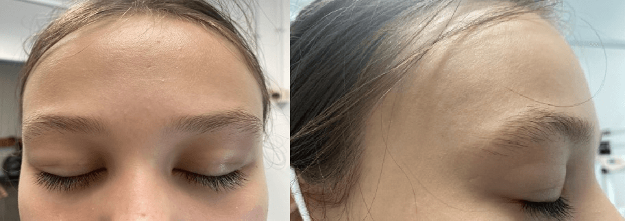 What is the diagnosis?
|
Discussion :
Upper eyelid edema as a manifestation of infectious mononucleosis (IM) was originally described by Colonel Robert J. Hoagland in 1952, hence the term “Hoagland’s sign”. Periorbital edema is a less common manifestation of IM, occurring in up to a third of patients. 1 Regarding the presentation of the sign, upper eyelid edema occurs before the onset of pharyngitis and cervical lymphadenopathy, but after the onset of fever. There is no associated proteinuria, eyelid inflammation or conjunctivitis and the eyelids are not tender. 2 The cause is unknown, but nasopharyngeal replication of the virus, lymphoproliferation or lymphatic obstruction are assumed to be the contributing factors. It is a helpful sign for distinguishing IM from other causes of viral pharyngitis or from streptococcal pharyngitis. 4 Bilateral periorbital edema in the absence of general edema can occur associated with other entities such as allergic reactions (angioedema), trichinosis, Kawasaki disease or bilateral periorbital cellulitis. 5
There are few published reports of Hoagland’s sign. This case emphasizes periorbital edema as an alerting symptom of EBV infection that physicians should keep in mind. | References : | - Hoagland RJ. Infectious mononucleosis. Am J Med. 1952; 13: 158-171.
- Feinberg AS, Spraul CW, Holden JT, et al. Conjunctival lymphocytic infiltrates associated with Epstein-Barr virus. Ophtalmology 2000; 107:159-63.
- Van Hasselt W, Schreuder RM, Houwerzijl EJ. Periorbital oedema. Neth J Med. 2009; 67: 338-339.
- Inokuchi R, Iida H, Ohta F, Nakajima S, Yahagi N. Hoagland sign. Emerg Med J. 2014; 31: 561.
- Suer KH, Kaptanoglu AF. Association of periorbital edema and fever in acute infectious mononucleosis: A case report. Kafkas J Med Sci 2013; 3: 152-154
|
|
| Correct Answers : |  100% 100% |
Last Shown : Apr 2025
|