Mariana Silva Duarte1, Filomena Pinto2, Rodrigo Carvalho3, Marta Conde4
1Medical Paediatric Unit, Área de Pediatria, Hospital de Dona Estefânia, Centro Hospitalar de Lisboa Central – EPE, Lisbon, Portugal, 2Neonatal Intensive Care Unit, Área de Pediatria, Maternidade Alfredo da Costa, Centro Hospitalar de Lisboa Central – EPE, Lisbon, Portugal, 3Dermatology Department, Hospital dos Capuchos, Centro Hospitalar de Lisboa Central – EPE, Lisbon, Portugal, 4Pediatric Rheumatology Unit, Área de Pediatria, Hospital de Dona Estefânia, Centro Hospitalar de Lisboa Central – EPE, Lisboa, Portugal
Address for Correspondence: Mariana Duarte, Rua Jacinta Marto, 1169-045 Lisboa, Portugal
Email: marianaduarte@campus.ul.pt
|
Question :We describe a case of a female newborn, from mother with systemic lupus erythematosus (SLE), with onset of skin lesions since day seven of life. Examination revealed six annular, erythematous, scaly patches on the face, trunk and lower extremities (Figures 1 and 2). Laboratory tests showed hemoglobin 9.6 g/dL, leukocytes 8.77 x10⁹/L, platelets 239x109/L, ANA >1/640 and positive extractable nuclear antigens (ENAs) (anti-Ro/SSA, anti-La/SSB and RNP/Sm). Cardiologic evaluation didn’t reveal anomalies. Skin biopsy showed apoptotic keratocytes with hydropic degeneration of the basal epidermis and perivascular lymphocytic infiltrate of the dermis compatible with neonatal lupus (NLE). Low potency topical corticosteroids (LPTC) were started.
Because of skin worsening (Figure 3), liver enzymes elevation (AST 225 U/L, ALT 87 U/L) and thrombocytopenia (115x109/L), low-dose systemic corticosteroids (SC) were started. Progressive reduction of SC until discontinuance was possible with normalization of the laboratory results and skin remission.
Figure 1. Scaly patches on the face. 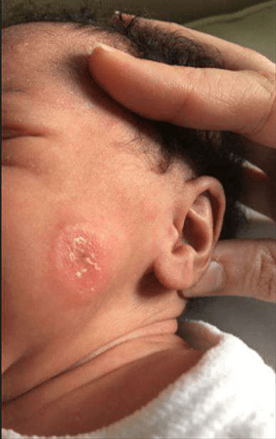 Figure 2.
 Scaly patches on the trunk and lower extremities. 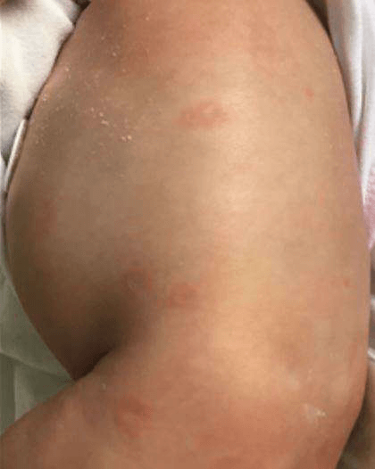 Figure 3.
 Skin worsening. 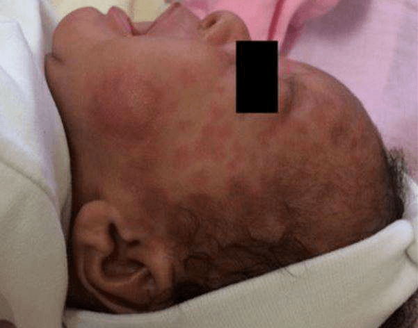 What is the diagnosis?
|
Discussion :
NLE is a rare autoimmune disease resulting from the passive transfer of maternal autoantibodies (MAA) (anti-Ro/SSA, anti-La/SSB and/or Anti-U1RNP) to fetus.
Congenital heart block may be detected in utero (2nd trimester) and is the classic cardiac manifestation, being determinant for prognosis because it is usually permanent and may require pacemaker placement.
Half of the patients present cutaneous manifestations that typically resolve until the first year of life (disappearance of MAA) and less frequently may present hepatobiliary and hematological involvement. 1,2
The diagnosis of NLE is based on clinical features and autoantibodies identification, but sometimes skin biopsy might be needed. Photoprotection and LPTC for short periods of time are the treatment of choice for cutaneous involvement, but SC can be used in patients with hepatobiliary and hematological manifestations. 1,3,4
Specially in cases without a previous diagnosis of the mother (40-60%), NLE diagnosis requires a high level of suspicion because cutaneous manifestations may mimic other common dermatosis. Even in mothers with previous SLE diagnosis, only 1-2% will have children with NLE (coexistence of Ac anti-Ro and anti-La increases this risk). Cardiological and laboratory evaluation is mandatory during pregnancy and after birth, allowing optimization of clinical care. 2,4| References : | - Teixeira AR, Rodrigues M, Guimarães H, Mura C, Brito I. Neonatal lupus - case series of a tertiary hospital. Acta Reumatol Port 2012; 42:318-23
- Izmirly PM, Halushka MK, Rosenberg AZ, Whekton S, Rais-Bahrami K, Nath DS, et all. Clinical and pathologic implications of extending the spectrum of maternal autoantibodies reactive with ribonucleoproteins associated with cutaneous and now cardiac neonatal lupus from SSA/Ro and SSB/La to U1RNP. Autoimmun Rev 2017; 16: 980-3
- Wisuthsarewong W, Soongswang J, Chantorn R. Neonatal Lupus erythematosus: clinical character, investigation and outcome. Pediatr Dermatol 2011; 28 (2): 115-21
- Hon KL, Leung AK. Neonatal lupus erythematosus. Autoimmune Dis. 2012; 2012:1-6
|
|
| Correct Answers : |  100% 100% |
Last Shown : Feb 2025
|