Dr Ira Shah.
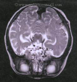
Medical Sciences Department, Pediatric Oncall, Mumbai, India. | | Case Report | A 10 month old female child (only sibling) born of non- consanguineous marriage presented with loose motions and vomiting since 3 days. She had 2 episodes of multifocal convulsions 1 month back for which she was admitted in a private hospital and treated with carbamazepine. A CT brain was done which showed periventricular calcifications. Her birth was uneventful with a birth weight of 3 kg. There was no history of rash or fever in the mother during pregnancy. Her milestones were slightly delayed (She achieved social smile at 5 months of age, head holding at 6 months of age, sitting with support at 10 months of age and babbling at 10 months of age). She was immunized till age and weaning had been started. On examination, she was well nourished (weight = 9.5 kg., 50th centile, head circumference = 44 cm, 50th centile and height = 74 cm, 50th centile) She had insignificant cervical lymphadenopathy. There were no neurocutaneous markers. On neurological examination, higher functions bone and power were normal though deep tendon reflexes were brisk. Other systemic examination revealed hepatosplenomegaly. In view of multifocal convulsions, hepatosplenomegaly and intracranial calcifications, congenital infection was suspected, A differential diagnosis of hyperparathyroidism and AICARDI CONTRI syndrome was also considered. Her TORCH titres revealed positive Cytomegalovirus (CMV) IgG [(95 IU/L (Normal = <10 IU/L)] whereas CMV IgM was negative. Her serum calcium, phosphorus, alkaline phosphatase were also normal. EEG was normal. Her ophthalmologic evaluation showed no chorioretinitis or cataract. Her liver profile and X-Ray chest were also normal. Her urine for CMV was negative. With persistent loose motions, she went into septic shock and was treated with IV antibiotics and ionotropic support. An MRI brain was done which showed extensive nodular calcifications in periventricular white matter with generalized cerebral atrophy suggestive of in-utero insult with perimesencephalic vasculopathy suggestive of CMV infection (Figure 1).
Figure 1 :MRI showing periventricular calcifications
The child continued to have multiple episodes of convulsions which required 3 anticonvulsants to control. The child then became lethargic and lost all previous milestones. Parents were counseled regarding ganciclovir, however was not given due to non affordability.
Thus, this is a case of a child who had delayed milestones and presents in late infancy with symptoms of intrauterine infection that leads to CNS manifestation later in life confirming that intrauterine infections may manifest after the para-neonatal period too. | | | | Discussion | CMV is the largest of the herpes viriolae group and incidence of congenital CMV infection in the western countries ranges from 0.2 to 2.4% of all live births. The risk for fetal infection is greatest with maternal primary CMV infection (30%) and much less likely with recurrent infection (<1%). Perinatal transmission has an incidence of 10-60% and can occur during the 1st 6 months of life with mode of transmission being the genital tract secretions at delivery and through breast milk.
Symptomatic congenital CMV infection was originally termed cytomegalic inclusion disease. (hepatosplenomegaly, jaundice, petechiae, purpura and microcephaly). Other features seen are intrauterine growth retardation, prematurity and intracranial calcifications. Other neurologic problems include chorioretinitis, sensorineural hearing loss. Only 5% of all congenitally infected infants have severe cytomegalic inclusion disease, 5% have mild involvement and 90% have subclinical but chronic CMV infection. Perinatally acquired CMV can lead to pneumonia and sepsis like syndrome, however most are asymptomatic.
Symptomatic congenital CMV has presentations:
- Early manifestation: Presents with involvement of multiple organ systems with a high risk of mortality. Findings include petechiae or purpura, hepatosplenomegaly, jaundice and "blue berry muffin spots" consistent with extra medullary hematopoiesis. Laboratory abnormalities include elevated hepatic transaminases, hyperbilirubinemia, anemia and thrombocytopenia.
- A second early presentation includes those infants who are symptomatic but without life threatening complications. These babies may have IUGR or microcephaly with or without intracranial calcifications. These calcifications are classically seen in the periventricular area as was seen in our case. Patients may have chorioretinitis, sensorineural deafness and developmental abnormalities.
Those with asymptomatic infection may have hearing loss, mental retardation, motor spasticity and microcephaly with abnormal enamel production later in life.
Diagnosis
Diagnosis is by viral isolation or PCR which should be performed at or shortly after birth in urine, saliva, blood or respiratory secretions. If found within first 2 weeks of life, it denotes congenital infection. If found after 4 weeks of life, it may denote perinatal infection. CMV antigen can be detected in blood. Positive results confirm CMV infection and viremia; however, negative results do not rule out CMV infection.
Maternal serum antibodies (CMV specific IgG) show a decline within a month and uninfected children have no detectable titer by 4 to 9 months. Infected infants continue to produce IgG throughout the same time period. Our patient had a high CMV IgG even at the age of 10 months suggestive of CMV infection in the child. IgM tests lack specificity and sensitivity for diagnosis of congenital CMV.
Treatment
Treatment by ganciclovir (6 mg/kg/dose IV 12 hourly) for first 6 weeks of life has demonstrated improved hearing and prevention of hearing deterioration though drug-related toxicity is common. Treatment with ganciclovir is still under evaluation. | | | | Compliance with Ethical Standards | | Funding None | | | | Conflict of Interest None | | |
- Behrman RE, Kliegman RM, Jenson HB. Nelson Textbook of Pediatrics. 17th ed, W.B.Saunders, Philadelphia, 2004;1066-1068.
- Cloherty SP, Eichenwald EC, Stark AR. Manual of Neonatal care. 8th ed. Lippincott Williams & Wilkins, Philadelphia. 2004; 255-259.
|
| Cite this article as: | | Shah I. CONGENITAL CYTOMEGALOVIRUS INFECTION - A LATE MANIFESTATION. Pediatr Oncall J. 2004;1. |
|